- Features
- Specification
- Application
- Promotional Video
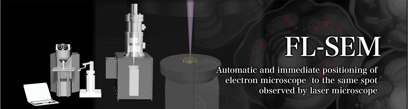
FL-SEM
FL-SEM is a correlation microscope system that combines the laser microscope performing fluorescence observation of the biological sample, electron microscope performing microstructural observation, and image processing technique. The greatest feature of the system is the possibility to immediately observe the place with the electron microscope, which was observed with the laser microscope. Until now the manual movement and alignment of the sample, in the mutual observation between the laser microscope and the electron microscope is not realistic. The correlative analysis reached the practical level for the first time, with the sample transportation system CAT's (Cooperation Automatic Transport System), the precision stage of this system, and by the unified management of the observation position in the laser microscope and the electron microscope. High definition fluorometric imaging is possible by the fluorescent observation that visualizes the physiological activity of cell, and the electron microscope observation that specifies cellular ultrastructure.
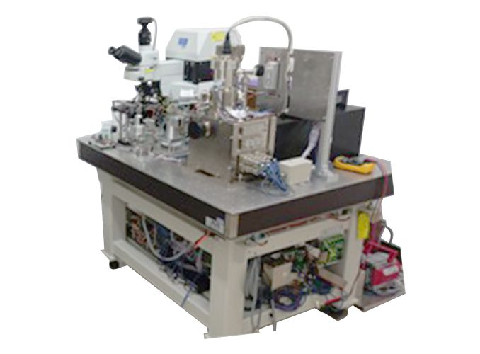
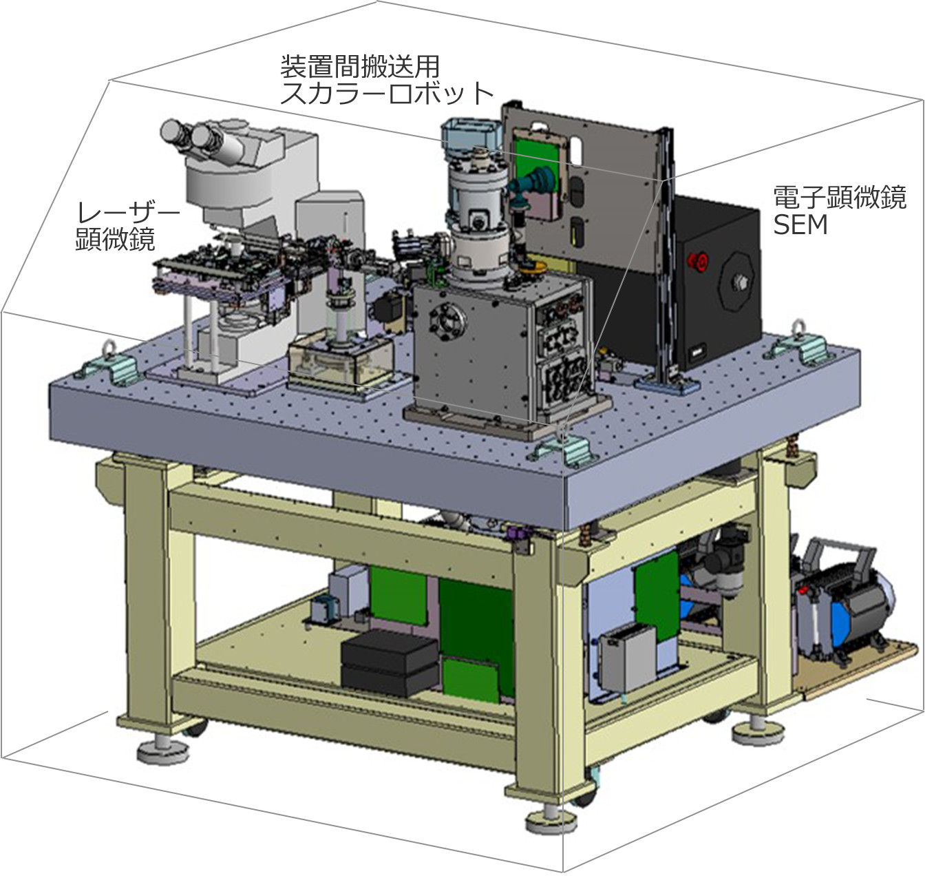
Configuration example of FL-SEM
The detail of FL-SEM configuration
This instrument is composed of SEM, specimen transporting unit, and unified management system that controls all units and modules.
Primary specification
| FL (Fluorescent Laser microscope) part | |
|---|---|
| Laser wavelength | This instruments is based on the laser microscope standard of various optical microscope makers. |
| Laser output | |
| Laser light source | |
| Magnification | |
| Sample size | |
| SEM (Scanning electron microscope) part | |
| Emitter | ZrO/W |
| Accelerating voltage | 1 - 5kV (continuous variable) |
| Resolution | 10nm |
| Magnification | x50 - x500,000 |
| Sample size | 0.5 inches |
| Stage components | X-Y-Z-R Automatic |
| Stage precision | Repeated positioning ± 0.1um |
| Detection of stage position | Linear encoder 0.01um |
| Exhaust system | Dry exhaust system |
| Sample conveying | Fully Automatic |
| CAT's -Automatic sample-conveying device- | |
| Fully automatic conveying system | |
Application
High definition fluorometric imaging in cancer diagnosis
The optical magnification in case of laser microscope unit is several 100 ~ several 1000 times, and it cannot specify which part of the organelle is fluorescently stained. However, it is possible to analyze the fluorescent staining analysis at the nanometer order level, by the electron microscope having the magnification rate of several tens of thousands of times and by the image synthesizing.
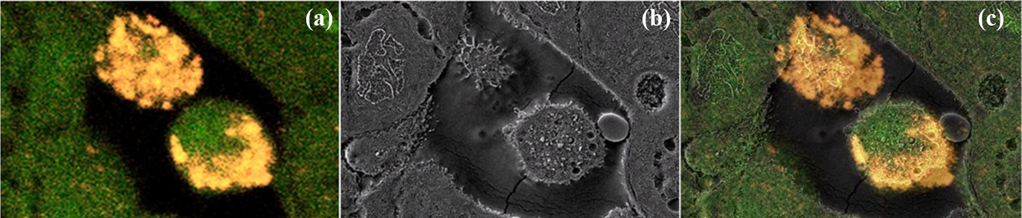
Left: Fluorescent image (Objective lens about 60 times), Center: Electron microscope images (about 3,000 times), Right: Image synthesized FL-SEM image
Operation principle of FL-SEM
This 1 minute movie explains the features and mechanism of FL-SEM.