Cell observation
Coming soon
Coming soon
Only with laser microscope, the magnification ratio is approx. 10 to 100 in objective lens. So we cannot distinguish which organelle is fluorescently dyed. But combining SEM image which magnification ratio is over x1,000m we can analyze the fluorescence in nano meter order.
| (a)FL image (Objective lens x60 approx.) |
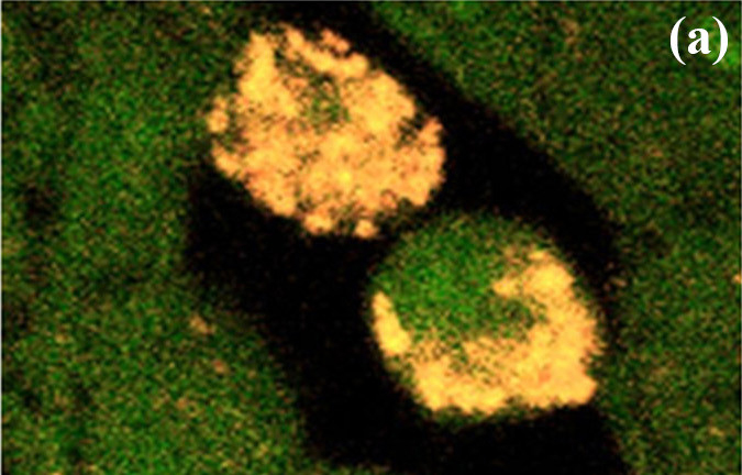 |
| (b)SEM image (x3,000 approx.) |
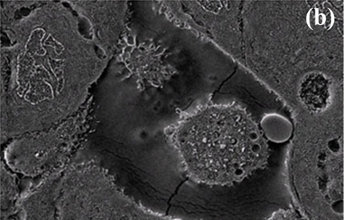 |
| (c)Fast processing image of FL-SEM |
 |
Coming soon
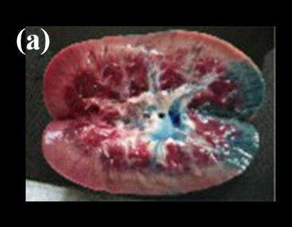
Nanometer order and high resolution 3D modeling of an experimental animal kidney is available by using our CT-SEM.
|
Left: Kidney coated by epoxy resin, Right: 3D image of kidney glomeruli by using CT-SEM
Patterning of resist of silicon wafer by using the electron beam (EB) attachment.
 |
| Small and all-in-one electron microscope with measurement and fabrication platform |
Photoresist used: ZEP520A (Xeon)
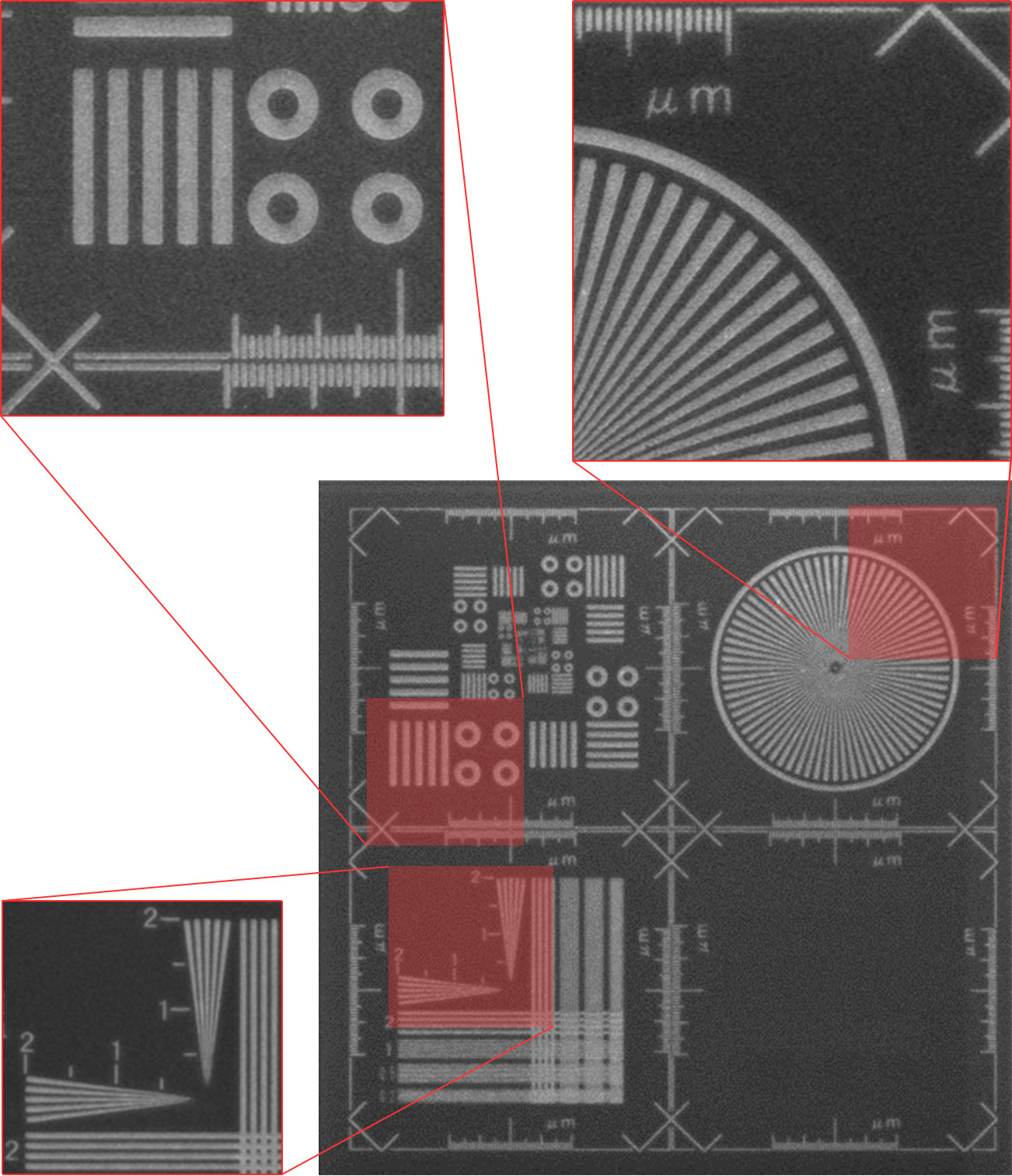
The size measurement of resist patterns by using minimal CD-SEM. Simple and easy measurement of fine device pattern is realized by using the software included.
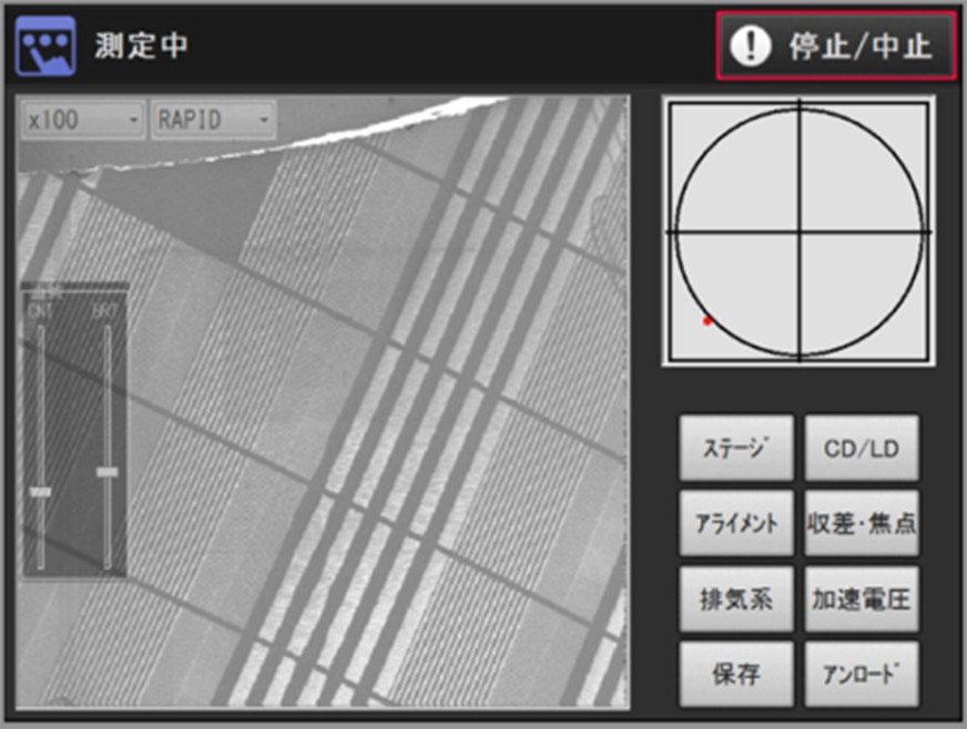
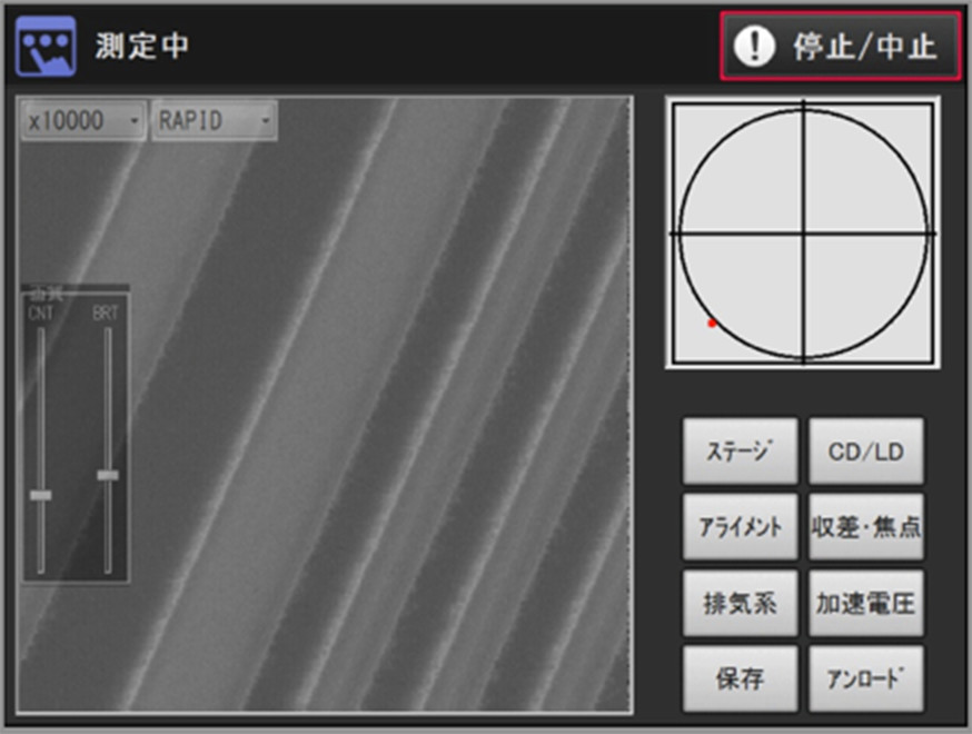
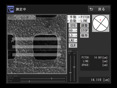
Screenshot of inclueded software. From left: x100, x10,000, processing screen
Coming soon
Coming soon
Coming soon
Coming soon
Coming soon
Coming soon
Coming soon
Coming soon
Coming soon
Coming soon